


Immuno-Histochemistry: Everything You Need to Know

February 19, 2026
Over recent years, advances in diagnostic pathology have substantially transitioned from total morphological evaluation of tissue to the molecular and immunological characterization of tissues. A significant contributor to this evolution has been the use of Immuno-Histochemistry (IHC). IHC is an established laboratory procedure that incorporates the principles of immunology, histology and biochemistry by detecting and localizing specific antigens found within tissue sections. The identification of protein expression patterns is accomplished through the use of antigen-antibody interactions visualized using chemical staining, while also preserving the three-dimensional architecture of the tissue.
What is Immuno-Histochemistry?
Immuno-histochemistry (IHC) is a laboratory method to determine, locate and visualize the position of a specific antigen, typically a protein found in a fixed tissue section. IHC is based on the principle of specific antigen-antibody interactions, where an antibody will specifically bind to its corresponding antigen, which may be present in the tissue or cell type being evaluated. Once an antibody binds to its specific antigen, IHC uses either enzymatic or fluorescent methods to identify the location of the target molecule, while still preserving tissue morphology, thus enabling the use of a microscope to identify the target molecule.
IHC combines the principles of immunology, histopathology, and chemical analysis of tissue. Unlike histological stains, which identify only the overall histological architecture of the tissue, IHC provides molecular specificity for identifying the cell lineage, functional status and any pathological changes at the level of protein expression. Some of the common applications of IHC are:
- Oncology
- Infectious Diseases
- Neuropathology
- Biomedical Research
How Does IHC Work?
Immunohistochemistry is based on the principle of specific binding of an antibody to a particular antigen, which allows for the specific identification of target proteins in tissue sections. The process of immuno-histochemistry begins with obtaining a tissue sample via either biopsy or surgical excision, with the subsequent fixation of the tissue sample to preserve cellular architecture and maintain the integrity of the antigen therein. The tissue sample is then prepared and cut into thin sections for subsequent microscopic examination.
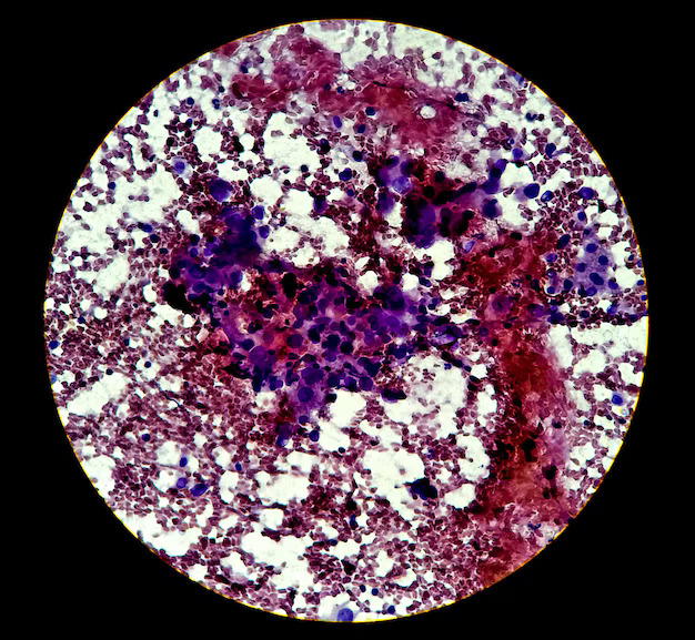
Once the tissue has been prepared, a primary antibody directed to the protein of interest is then applied directly to the tissue section. In the event that the target protein is present, the antibody will bind to the antigen, forming an antigen-antibody complex. Antigen-antibody complexes are visualized using either an enzymatic or chromogenic reaction to produce a coloured precipitate, most commonly a brown colour, at the location of the target protein in the tissue. The resultant staining pattern aids the pathologist in determining the localization and relative abundance of the target protein, thus assisting in accurate diagnosis and interpretation.
Limitations of Immuno-Histochemistry
IHC has a diagnostic reason for being valuable, but it also has limitations. The reproducibility of IHC is primarily contingent upon the individual antibody being used, its availability, and the final product being produced by that antibody. There is variability from one batch of antibodies to another; the final result will also vary from batch to batch, depending on the individual performance of each antibody’s production.
- The interpretation of staining patterns associated with an IHC study is often subjective in nature and therefore requires experience and expertise in pathologic interpretation.
- The interpretation of staining patterns is subjective, and polymeric handlers of stained tissue impose their individual expertise on tissue staining patterns when looking at IHC.
- Tissue fixation, antigen retrieval, and the protocols used to perform antibody staining all contribute to the variability in the technical aspects of IHC processing.
- Variability in laboratory processes and procedures can affect the reproducibility and comparability of the final stained tissue specimen produced by the laboratory.
- IHC is a capital-intensive laboratory procedure that requires specialized laboratory equipment and trained laboratory personnel be available in order to successfully carry out an accurate IHC processing procedure.
Conclusion
Immuno Histochemistry (IHC) has become a fundamental part of modern-day diagnostic pathology, allowing us to identify and locate specific proteins within tissues with precision. By using the combined strength of specificity from immunology while simultaneously retaining the morphology of the tissue, IHC allows one to make accurate diagnosis, classify diseases, prognosticate, and make therapeutic decisions.
For DPMI, IHC is a foundational area of study for future health care professionals. DPMI staff and students work to develop their comprehension of IHC principles, their IHC work in the laboratory, and its education of clinical application. Ongoing training, exposure to hands-on activity, and higher level of academic involvement related to advanced diagnostic techniques such as IHC will provide paramedical professionals with the tools necessary to advance in the continually changing landscape of 21st century medicine.

