


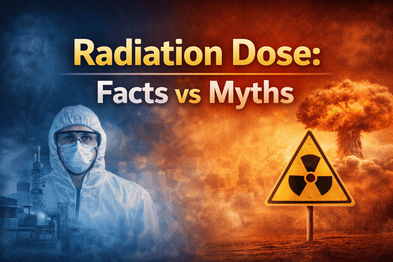
Radiation Dose: Facts Vs Myths

February 11, 2026
Till date, a huge chunk of people are fearful of medical radiation and often base that fear on misconceptions rather than an understanding of science. Despite the extensive use of X-rays, CT scans, and other imaging techniques, myths and misunderstandings about radiation dose and associated risks persist. These misunderstandings frequently create unnecessary anxiety for patients, leading them to refuse required diagnostic imaging and perpetuate misinformation about medical radiation.
There is an established set of safety standards for the use of medical radiation, as well as strict protocols to monitor the amount of radiation to which patients are exposed, all backed by internationally accepted dosimetry standards. Because medical radiation is tightly regulated, it is critical to understand what a radiation dose is and how radiation dose is measured, along with a historical perspective on the management of patient exposure through new technologies. The objective of this blog is to establish a framework that bridges the gap between perception and evidence regarding medical radiation, facilitating a comprehensive understanding of both established scientific facts and common myths surrounding it.
What is a Radiation Dose in the Medical Field?
Radiation dose in the healthcare setting is defined as the amount of ionising radiation energy absorbed by the body's tissues during imaging or treatment procedures. The term “radiation dose” is a quantitative measure of exposure and is used to assess the benefits and risks of using imaging techniques such as X-rays, CT scans, fluoroscopic procedures, and nuclear medicine.
Radiation doses are described in millisieverts (mSv), which not only account for the physical energy absorbed by the tissue but also for how that absorbed energy will affect the biological systems of the body. The reason for this distinction is that various forms of ionising radiation, such as X-rays, gamma-rays, and beta particles, produce different effects on living cells, even though they are all absorbed at the same energy level.
Facts vs Myths About Radiation Dose
There is a large number of misconceptions regarding medical radiation that exist today. Many of these are the result of incomplete and outdated information or the general fears associated with experiencing radiation exposure. The result is that patients tend to overestimate the risks related to using diagnostic imaging and underestimate the protective mechanisms inherent to modern radiology. Patients need to know what scientifically validated information is available so they can make informed decisions about their use of imaging technologies and have greater assurance when utilising these imaging technologies. By providing a comparison to common misunderstandings, this section aims to clear up some of the common myths surrounding medical radiation.
Myth 1: Any amount of radiation is dangerous.
Fact: The myth of “Radiation is dangerous at all levels” has been around for years. Radiation is a naturally existing phenomenon within our environment and is present as UV rays from the sun, Soil and Food, in addition to being produced in our bodies. The average person receives an estimated annual background radiation exposure of 2-3 mSv. The additional radiation exposure from medical imaging occurs in a controlled manner and presents minimal additional exposure compared to the overall benefits obtained.
Myth 2: CT scans expose you to dangerously high radiation.
Fact: While it is true that CT scans do have a higher radiation dose than standard radiographs, the actual radiation doses are still within acceptable limits. For instance, a standard abdominal CT scan may be 5-8 mSv, which is equivalent to the amount of radiation the average person will receive from natural background radiation over a period of 2-3 years.
In addition, advancements in CT technology permit the use of dose optimisation techniques to maintain the patient's exposure to radiation at a level that is reasonably possible to keep to a minimum.
Myth 3: Medical radiation always increases cancer risk.
Fact: The relationship between low doses of medical imaging and cancer risk is often misreported. The increases in cancer risks due to the levels of radiation associated with medical imaging, in particular the levels associated with X-rays, Mammograms, Dental, Low-dose CT scans, etc., are incredibly small, if they are detectable, and therefore statistically inconclusive in comparison to levels of natural environmental exposure for most people. Therefore, the diagnostic positive benefits from imaging versus the risk of diagnosis are extremely beneficial to the patients.
Myth 4: Pregnant women must never undergo imaging
Fact: Ultrasound and MRI are safe to use during pregnancy since they do not emit ionising radiation. X-ray diagnostic imaging, such as for dental work or in the case of chest X-rays, involves such low exposure of the fetus as a result of the manufacturer’s shields and careful imaging methods. In cases of emergency, X-rays or imaging techniques may be necessary to ensure that both the mother and her unborn child are safe.
Myth 5: Lead aprons eliminate radiation exposure.
Fact: While lead aprons are intended primarily to protect against scatter radiation, they do not eliminate a patient’s exposure to the primary beam. Lead aprons do provide an important measure of protection against scattered radiation in certain circumstances. However, they do not protect the patient from direct exposure to ionising radiation. Shielding in the past served as the primary method of protecting patients from direct exposure. However, it is now used mainly as supplementary protection rather than as a primary method of protecting patients.
Conclusion
To safely and effectively utilise medical imaging, it is critical that patients, health care providers, and all individuals working with medical imaging are educated on the distinctions between “fact-based evidence” obtained from studies and the common myths surrounding radiation dose. Today, because of the stringent safety requirements imposed by regulators and health care institutions, the use of medical imaging is more thorough, accurate, and reliable than ever before. By utilising accurate sources of information, both patients and health care providers will be able to make informed diagnostic decisions more quickly and accurately.
For those aspiring to build a career in this field, comprehensive radiology tech courses provide the essential knowledge of radiation safety, imaging physics, and patient care required in today’s healthcare environment. Institutions that emphasise practical training and scientific clarity play a pivotal role in preparing skilled professionals.
If you want to pursue a rewarding career in medical imaging, DPMI offers industry-aligned radiology programs designed to equip you with the right skills, hands-on experience, and professional competence. Enrol with DPMI today and take the first step towards securing a safe medical career.

